

We didn’t notice the ‘issue’ until he was maybe six months old. We’ve asked our vet about it twice. It’s a larger practice so we’ve had two opinions over the course of approximately 1 year.
At his first annual exam the vet didn’t really see anything wrong. She referred to it as “bright” in her veterinarian note. We recently brought him into for is annual exam (approx 1 year later) and this time we saw a different vet. She actually brought it to our attention.
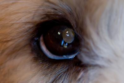
We discussed it at some detail. However, she wasn’t sure what it really was. She just said that there was a “change in the lens of the right eye.” She recommended a consult with a veterinary ophthalmologist.
The eye doesn’t seem to be bothering him at all, and I haven’t noticed it getting any worse since we first spotted it.
It’s quite noticeable in the right lighting conditions. I’m hoping my picture is detailed enough. I have a much higher resolution image that provides way more detail than the upload tool will accept. Please let me know if you would like me to email this copy.
Thanks for your assistance – we are anxious to determine the issue and what can be done if anything.
Dog’s Age: 2
Gender: Male
Breed: Soft Coated Wheaten Terrier
Comments for Dog Eye Condition? | ||
| ||
Click here to go back to the Ask a Vet Online Library of questions.

Do you believe in holistic pet care? If so, please tell your friends about us. Thank you for supporting our efforts!
Also see…
- Back to Dog Health Problems Symptoms / Dog Illness Signs Symptoms / Natural Dog Remedies
- Back to 10 Best Dog Food Options / Dog Food Ratings / Buy Dog Food Online
- Back to Conventional vs. Holistic Veterinarians
- Back to Organic Dog Supplies Online
- Back to Pet Friendly Airlines / Pet Friendly Travel
- Back to Organic Pet Digest Natural Dog Care Home Page